

Nanobody-fluorescent protein fusions for erasable immunostaining—Xinxin Xu; Zhe Kong; Zhipeng Su; Chen Cheng; Yuan Wan
Abstract
Immunofluorescence (IF) staining is widely used to visualize biomolecules in cells and tissues. Conventional IF typically relies on fluorophore-conjugated antibodies, whose preparation requires chemical modification procedures that can compromise antibody performance and incur product loss during subsequent purification. In addition, iterative immunostaining depends on harsh stripping or fluorescence-quenching treatments that may damage sample architecture and epitope integrity. Here, we present protease-cleavable nanobody-fluorescent protein fusions (NFPFs) for reversible IF staining. Unlike fluorophore-labeled antibodies, NFPFs are genetically encoded and can be produced without post-synthetic chemical modification. By incorporating a tobacco etch virus (TEV) protease cleavage site between the nanobody and fluorescent protein, we engineered HER2- and EGFR-targeting NFPFs that retained high target specificity and enabled robust, concentration-dependent immunostaining of Capan-1 cells. TEV-mediated cleavage efficiently removed the fluorescent reporter while preserving cellular morphology and antigen accessibility, allowing subsequent staining of targets. Sequential labeling of distinct receptors demonstrated the feasibility of iterative immunostaining within the same specimen. Collectively, NFPFs provide a simple, scalable, and versatile approach for molecular imaging, enabling repeated rounds of immunostaining.
Visual Abstract
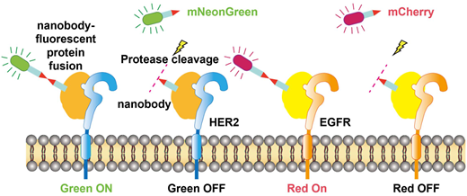
Protease-cleavable nanobody-fluorescent protein fusions can be produced by cell factories without post-synthetic chemical modification and enable reversible immunofluorescence staining.
1. Introduction
Fluorophore-labeled antibodies are widely used in immunofluorescence (IF) staining to visualize the distribution and expression of biomolecules within cells and tissues. Their preparation typically relies on covalent conjugation. However, chemical labeling can impair antigen-binding capacity and yield variable conjugation efficiencies.1–3 In addition, this labor-intensive modification and purification process compromises final antibody yields, driving up overall production costs.4 While covalent conjugation ensures stable fluorophore attachment to antibodies, it also poses a challenge for multiplexed IF imaging. In most fluorescence microscopies, the number of spectrally resolvable channels is limited to three or four, restricting the number of biomarkers that can be interrogated within a single staining round. Consequently, achieving higher levels of multiplexing generally requires iterative rounds of staining, signal erasure, and re-staining. Current signal-removal strategies, such as antibody elution using harsh reducing or acidic conditions and fluorophore quenching by oxidative bleaching, frequently damage tissue morphology and impair epitope integrity.5 These deleterious effects compromise staining fidelity and limit the depth of multiplexed molecular profiling.6 Therefore, developing a controllable, non-destructive method for fluorescence signal removal remains essential. Recent advances, such as CODEX and Exchange-PAINT, have expanded multiplexing capacity, enabling the profiling of dozens of molecular targets within a single specimen.7–9 These approaches rely on antibodies conjugated to DNA barcodes or docking stands, which undergo iterative cycles of fluorescent probe hybridization, imaging, and probe de-hybridization to achieve multiplexed molecular imaging. Despite their powerful multiplexing capability, both approaches require extensive optimization of DNA oligonucleotide design, hybridization conditions, and probe-stripping workflows.10 Moreover, the generation of DNA-conjugated antibodies still relies on chemical modification, thereby inheriting many of the aforementioned limitations associated with fluorophore–antibody labeling. Alternatively, chemically cleavable and photocleavable antibody–fluorophore conjugates enable the physical removal of fluorescence reporters through linker cleavage.11,12 In addition to antibody, nanobody i.e., NanoPlex, has been used in erasable fluorescence imaging platforms. These nanobody-based probes support iterative fluorescence imaging through photochemical, enzymatic, or chemically cleavable reporter modules. Of note, this technique uses primary antibodies to recognize targets, followed by binding with nanobodies modified with removable fluorescent reporters. Collectively, despite advances in erasable-labeling and iterative imaging technologies, further improvements are needed in molecular design, the efficiency and controllability of signal removal, and the overall simplicity of experimental workflows.13,14 Accordingly, we investigate nanobody-fluorescent protein fusions (NFPFs). Each NFPF integrates a target-specific nanobody, a tobacco etch virus (TEV) protease recognition site, and a fluorescent protein within a single recombinant polypeptide. This architecture enables the staining reagent to be produced without post-expression fluorophore conjugation or DNA-barcode installation, while TEV-mediated cleavage provides a sequence-specific mechanism for removing the fluorescent reporter after imaging. The principal distinction of this approach therefore lies not in the general concept of iterative staining, but in the use of a directly expressed, protease-cleavable NFPF as the staining reagent. In this study, we generated HER2- and EGFR-targeting NFPFs and evaluated their recombinant production, target-binding properties, cellular staining, TEV-mediated fluorescence removal, and applicability to sequential immunostaining.
2. Experimental sections
2.1. Cell culture
HEK293 cells and Capan-1 cells were cultured using Dulbecco's modified Eagle medium (DMEM; Corning, 10-013-CM) and RPMI-1640 medium (Corning, 10-040-LX), respectively, supplemented with 10% fetal bovine serum (FBS; VWR, 104026-678) and 1% penicillin–streptomycin. Polyethyleneimine (PEI; VWR, 76836-662), phosphate-buffered saline (PBS; VWR, 77975-369), bovine serum albumin (BSA; VWR, 77510-176), paraformaldehyde (PFA; VWR, 101176-014), and TEV protease were obtained from commercial suppliers. All reagents were of analytical grade and used as received unless otherwise specified.
2.2. Expression and purification of NFPFs
The NFPFs were designed with an N-to-C-terminal architecture consisting of a target-specific nanobody, a flexible linker, the TEV protease recognition sequence ENLYFQ↓S, a second flexible linker, a fluorescent protein, and a C-terminal His tag. The anti-HER2 nanobody (2Rs15d) was fused to mNeonGreen to generate HER2-mNeonGreen, whereas the anti-EGFR nanobody (7D12) was fused to mCherry to generate EGFR-mCherry.15,16 The complete amino-acid sequences of both constructs are provided in Table S1. NFPFs were transiently expressed in HEK293 cells. Plasmid DNA and PEI were premixed in serum-free medium for 20–30 min before transfection. Cells were maintained at 37 °C under 5% CO2, and culture supernatants were collected 72 h after transfection. NFPFs were produced using ten T225 flasks, which involved ten separate transfections. The resulting culture supernatants were pooled, yielding a total of approximately 300 mL of supernatant for subsequent NFPF purification. Secreted His-tagged NFPFs were purified by Ni-NTA affinity chromatography, dialyzed against PBS, and concentrated by centrifugal ultrafiltration. For prokaryotic expression, E. coli BL21 Star (DE3) cells were transformed with the corresponding plasmids. Protein expression was induced with 0.5 mM IPTG when the optical density at 600 nm reached approximately 0.7. Proteins were subsequently harvested and characterized.
2.3. SDS-PAGE analysis
NFPFs were analyzed by SDS-PAGE under non-reducing conditions. Approximately 20 µg proteins per lane was mixed with Laemmli sample buffer without reducing agents. Samples were heated at 70 °C for 10 min and subsequently loaded onto a 15% resolving polyacrylamide gel with a 4–5% stacking gel. A prestained protein molecular-weight marker was included for estimation of apparent molecular weights. Electrophoresis was performed using 1× Tris-glycine-SDS running buffer containing 25 mM Tris, 192 mM glycine, and 0.1% SDS. Gels were run at a constant voltage of 200 V for 40 min. Following electrophoresis, gels were removed from the cassette and stained with Coomassie Brilliant Blue for ∼1 h at room temperature with gentle agitation. The gels were subsequently destained until a clear background, and well-resolved protein bads were obtained and were imaging using a gel documentation system. Proteins were incubated with TEV protease at enzyme concentrations of 0, 1.5, and 2.5 U μg−1 for 0–4 h at 4 °C or 30 °C. Cleavage products were visualized by Coomassie Brilliant Blue staining.
2.4. Size-exclusion chromatography (SEC)
Purified NFPFs were characterized by size-exclusion chromatography using an Agilent 1260 Infinity II system equipped with an Agilent AdvanceBio SEC 300 column (2.7 μm, 4.6 × 300 mm). The column was equilibrated with 150 mM PBS (pH 7.0), which was also used as the mobile phase. Protein samples were clarified prior to injection and 5 μl of each sample was injected onto the column. Separation was carried out isocratically at a flow rate of 0.35 ml min−1 and a column temperature of 25 °C for 15 min. Protein elution was monitored by UV absorbance at 220 nm, and chromatograms were recorded and analyzed using the Agilent chromatography data system. The relative proportions of the main protein peak, aggregated species, and low-molecular-weight fragments were determined by chromatographic peak-area integration.
2.5. Enzyme-linked immunosorbent assay (ELISA)
Binding of NFPFs to their corresponding recombinant target proteins was evaluated using an immobilized-antigen ELISA. Recombinant human HER2 or EGFR was diluted in carbonate–bicarbonate coating buffer (pH 9.6) to 2 µg ml−1, and 100 µl was added to each well of Corning high-binding 96-well microplates. Please were incubated overnight at 4 °C to allow antigen immobilization. The coating solution was removed, and the wells were washed thrice with PBS containing 0.055 Tween-20 (PBST). Nonspecific binding sites were blocked with 3% BSA in PBS for 1 h at room temperature. NFPFs were prepared in PBS containing 1% BSA, yielding final concentrations of 0.000128–2 μg ml−1. Aliquots of 100 μl were added to the antigen-coated wells and incubated for 1 h at room temperature. Plates were subsequently washed thrice with PBST, and bound NFPFs were detected by incubation with an anti-His primary antibody for 1 h at room temperature, followed by an appropriate HRP-conjugated secondary antibody for 1 h at room temperature. After extensive washing with PBST, 100 μl of TMB substrate was added to each well and allowed to develop for 10 min in the dark. The reaction was terminated by adding 50 μl of 2 M H2SO4, and absorbance was measured at 450 nm using a microplate reader. Background absorbance from antigen-coated wells without NFPF was subtracted from the corresponding measurements. Each concentration was analyzed in replicate, and concentration-response curves were fitted using a four-parameter logistic nonlinear regression model to determine the apparent EC50 values.
2.6. Bio-layer interferometry (BLI)
Binding kinetics of the NFPFs were characterized by bio-layer interferometry. Biotinylated recombinant human HER2 or EGFR extracellular domains (ACROBiosystems) were immobilized onto streptavidin-coated biosensors, and HER2-mNeonGreen or EGFR-mCherry was used as the soluble analyte, respectively. Measurements were performed at room temperature in PBS supplemented with 0.02% Tween–20 and 0.1% BSA. In the initial measurements, NFPFs were analyzed at concentrations of 12.5, 25, 50, 100, and 200 nM. Association and dissociation were monitored for 300 and 600 s, respectively. Reference responses obtained from antigen-loaded sensors incubated in assay buffer alone were subtracted from the corresponding binding curves. Sensorgrams were fitted using a 1 : 1 Langmuir binding model to obtain the association rate constant (kon), dissociation rate constant (koff), and equilibrium dissociation constant (KD). The equilibrium dissociation constant was calculated as KD = koff/kon.
2.7. Immunostaining and fluorescence imaging
Cells were seeded into imaging dishes and cultured to approximately 70–80% confluency. Cells were fixed with 4% PFA for 15 min and washed three times with PBS. Non-specific binding sites were blocked using 3% BSA before staining. Fixed Capan-1 cells were incubated with HER2-mNeonGreen (0.01–1 μg mL−1) or EGFR-mCherry (1–10 μg mL−1) at 4 °C for various durations. Following staining and PBS washing, fluorescence and bright-field images were acquired using a Nikon Diaphot 300 PSM-2120 inverted phase-contrast microscope (10× objective) equipped with a Micros Austria 10×/0.25 NA Infinity Plan Achromatic objective and an AmScope MD310C-BS digital camera (3.1 MP color CMOS, USB 2.0, C-mount). Bright-field and fluorescence images were acquired to monitor NFPF-mediated cellular staining and subsequent fluorescence removal following TEV protease treatment. Hoechst nuclear fluorescence was additionally recorded in the experiments in which a nuclear reference was required. Fluorescence images were acquired using the corresponding Nikon fluorescence filter sets with an exposure time of 100 ms. For all samples subjected to direct quantitative comparison within an experiment, illumination, filter configuration, camera settings, exposure time, and other acquisition parameters were maintained unchanged. Fluorescence intensities were quantified using ImageJ using the same analysis procedure for all directly compared groups. Unless otherwise stated, repeated functional measurements were performed using aliquots from the same NFPF production batch; independently expressed and purified protein batches were not used as biological replicates. To evaluate nonspecific cellular association of the fluorescent proteins, fixed Capan-1 cells were incubated with free GFP (Abcam, ab84191) or mCherry (Abcam, ab199750). Cells were incubated for 1, 2, or 4 h using the same blocking, washing, and fluorescence-imaging workflow as that used for NFPF staining.
2.8. TEV protease-mediated fluorescence removal
NFPF cleavage was performed using TEV protease in 1× PBS. TEV activity was expressed as U μg−1 of NFPF, with the denominator referring to the mass of fusion-protein substrate in the reaction. The fusion constructs contained the TEV recognition sequence ENLYFQ↓S between the nanobody and fluorescent-protein domains. Cleavage was initially optimized at 30 °C using 0, 1.5, and 2.5 U μg−1 TEV over 0–4 h. Near-complete cleavage was observed at 2.5 U μg−1 after ∼2 h. Cleavage was subsequently evaluated at 4 °C using extended incubation periods to identify conditions more compatible with the cell-based staining workflow. Cleavage products were characterized by non-reducing SDS-PAGE.
2.9. Photobleaching measurement
A photobleaching experiment was performed under the same fluorescence acquisition conditions. NFPF-stained cells were imaged with an exposure time of 100 ms, with one image acquired every 500 ms for 20 consecutive acquisitions, corresponding to a total observation period of 10 s.
2.10. Statistical analysis
Statistical analyses were performed using GraphPad Prism. For cellular fluorescence measurements, each data point represents the mean fluorescence intensity measured from a segmented cell-associated region within one field of view obtained from one independently processed well. Unless otherwise indicated, five separate wells from the same cell preparation were analyzed per condition. For comparisons involving three or more independent groups, one-way ANOVA test followed by Dunnett's T3 multiple-comparisons test was used. Data are presented as mean ± SD, with individual well-level measurements displayed where appropriate. Statistical significance was defined as P < 0.05. ELISA concentration-response data were analyzed separately using four-parameter logistic regression. Two independent ELISA experiments were performed, each containing technical duplicate measurements at each concentration, and each independent experiment was fitted separately.
3. Results and discussion
3.1. Expression and characterization of NFPFs
The NFPF is organized in an N-to-C terminal orientation, comprising the NB, an upstream flexible (G4S)2 linker, a central cleavage motif, a downstream flexible (G4S)2 linker, and the fluorescent protein (Fig. 1a). Following the design, the anti-HER2 NB was genetically fused to mNeonGreen (HER2-mNeonGreen), while the anti-EGFR NB was fused to mCherry (EGFR-mCherry). To minimize steric hindrance and ensure efficient accessibility of the cleavable motif to the TEV protease, we incorporated (G4S)2 flexible linkers. Given the relatively compact sizes of the NB (∼16 kDa) and the fluorescent protein (∼27 kDa), a (G4S)2 unit, ∼3 nm in length, was sufficient to ensure efficient proteolytic cleavage. We did not choose a rigid linker EAAAK. EAAAK linkers primarily function as rigid α-helical spacers that constrain the relative distance and orientation of the connected domains. Such rigidity is beneficial when defined spatial separation between domains is required, but was not necessary for the present construct, in which the preservation of nanobody accessibility and independent fluorescence was prioritized.17 Both constructs showed a final molecular weight of ∼46 kDa. We evaluated both prokaryotic and eukaryotic expression systems for the production of the two NFPF constructs. The NFPFs produced in E. coli exhibited EC50 of ∼1.89 μg ml−1 for EGFR-mCherry and 2.85 μg ml−1 for HER2-mNeonGreen, respectively, indicating a significantly reduced binding affinity toward their targets compared to the HEK293-derived counterparts, which showed EC50 of ∼0.01 μg ml−1 for EGFR-mCherry and 0.027 μg ml−1 for HER2-mNeonGreen (Fig. 1b). We speculate that this discrepancy arises from suboptimal protein folding during synthesis in E. coli, which likely impairs the structural integrity and binding efficacy of the NB moieties.18,19 Given the performance and high yield of the NFPFs obtained from the HEK293 expression system, we opted to forego further optimization of the E. coli platform, such as exploring alternative strains, modulating induction temperatures, or fine-tuning IPTG (isopropyl β-d-1-thiogalactopyranoside) concentrations.20 All subsequent experiments were performed using HEK293-expressed NFPFs. Transient expression in HEK293 cells yielded 1.53 mg ml−1 of HER2-mNeonGreen at 87.39% purity and 10.3 mg ml−1 of EGFR-mCherry at 88.87% purity from the culture supernatant (Fig. 1c), as determined by size-exclusion chromatography (SEC). The binding properties of the HEK293-derived NFPFs were further characterized by BLI (Fig. 1d). Both NFPFs exhibited clear concentration-dependent association and dissociation kinetics. Global fitting using a 1 : 1 kinetic model yielded equilibrium dissociation constants of 1.85 nM for HER2-mNeonGreen and 1.7 nM for EGFR-mCherry. The error values for the Kd measurements of HER2-mNeonGreen and EGFR-mCherry are 2.1 × 10⁻¹¹ nM and 1.7 × 10⁻¹¹ nM, respectively. The corresponding association rate constants were 5.35 × 10⁵ M−1 s−1 and 5.94 × 10⁵ M−1 s−1, whereas the dissociation rate constants were 9.89 × 10⁻⁴ s−1 and 1.01 × 10⁻³ s−1, respectively. The Kd value of the two NFPFs were comparable to those previously reported for anti-EGFR nanobody 7D12 (3.6 nM) and anti-HER2 nanobody 2Rs15d (2.7 nM), indicating that fusion of the linkers and fluorescent protein to the C-terminus of the nanobody did not compromise target recognition or binding affinity.21,22 It should be noted that the measured Kd values may vary slightly depending on the assay method used.23 We next evaluated TEV protease cleavage to separate the NB and fluorescent protein domains. When the raw proteins harvested from the supernatant were analyzed by SDS-PAGE under non-reducing conditions, the NFPFs at ∼46 kDa were readily observable; however, additional bands were also present, indicating potential self-aggregation or partial degradation (Fig. 1e). For both NFPFs, cleavage efficiency exhibited a positive correlation with both TEV protease dosage and incubation time. Specifically, treatment with 2.5 U µg−1 of TEV protease effectively cleaved the NFPFs within 2 h at 30 °C, yielding liberated NB and fluorescent protein domains that strictly matched their expected molecular weights. Reducing the incubation temperature to 4 °C substantially slowed TEV-mediated cleavage, requiring 3 h to achieve near-complete processing. Notably, this cleavage efficiency depends not only on temperature and time, but also on TEV protease dosage.24 At a constant temperature, increasing the enzymatic load can compress the cleavage timeline.25 Together, these results demonstrate that the HEK293-expressed NFPFs retain high-affinity target recognition while permitting efficient TEV protease-mediated removal of the fluorescent protein domain, thereby enabling reversible immunostaining and sequential imaging.
Fig. 1
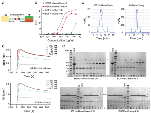
Design, expression, and functional characterization of two NFPFs. (a) Schematic illustration of the molecular architecture of the two NFPFs. NB: nanobody, and FP: fluorescent protein. (b) ELISA analysis comparing the target-binding capacities of NFPFs expressed in prokaryotic and eukaryotic systems. Each data point represents the mean of duplicate technical measurements. (c) SEC purity analysis of expressed NFPFs. (d) BLI sensorgrams detailing the concentration-dependent association and dissociation kinetics of the two NFPFs. (e) SDS-PAGE analysis of NFPF cleavage under varying TEV protease dosages (0–2.5 U) and incubation times (0–4 h) at different temperatures (4 °C and 30 °C).
3.2. Optimization of immunostaining and fluorescence removal
Immunostaining of the pancreatic adenocarcinoma cell line Capan-1 was performed using the NFPFs, respectively. Although Capan-1 cells concurrently express both HER2 and EGFR, their absolute expression levels are decoupled. HER2 is highly to moderately expressed, reported at a ∼5-fold higher density than EGFR, whereas EGFR demonstrates only moderate expression.26 Of note, some studies have also reported that the expression levels of both are very close, both at the level of ∼2 × 10⁴ per cell. Compounding this disparity, the intrinsic brightness of mNeonGreen exceeds that of mCherry by ∼4-fold.27 These varying observations are likely due to differences in the specific assay methods used for quantification. To account for these inherent biological and biophysical constraints, we initially sought to optimize the operational concentrations of NFPFs. At ∼70% confluency, incubation with 1 μg ml−1 HER2-mNeonGreen at 4 °C for 16 h produced robust labeling of virtually all cells, with a mean fluorescence intensity of ∼9 a.u. By comparison, 10 µg ml−1 EGFR-mCherry generated a lower mean fluorescence intensity of ∼2.5 a.u. (Fig. 2a). These differences are in agreement with the relative abundance of HER2 and EGFR in Capan-1 cells and the substantially higher brightness of mNeonGreen relative to mCherry. Subsequently, Capan-1 cells were incubated with 1 µg ml−1 of HER2-mNeonGreen and 10 µg ml−1 of EGFR-mCherry at 4 °C. For both NFPF constructs, we observed a progressive, time-dependent enhancement in the detected fluorescence intensities, characterizing the continuous binding of the NFPFs over the prolonged incubation window (Fig. 2b). Therefore, in subsequent experiments immunostaining was performed by incubating the cells with NFPFs overnight. In additional control experiments, incubation of free fluorescent proteins with fixed cells for up to 4 hours yielded no detectable fluorescence, demonstrating that these proteins do not non-specifically bind to the cell surface (Fig. S1). Subsequently, cells labeled with HER2-mNeonGreen were used to optimize the cell-surface cleavage efficiency of TEV protease, owing to its robust brightness and detection sensitivity. Capan-1 cells were treated with 2.5-, 3.75- and 5-U µg−1 of TEV protease at 30 °C for 2 h. This enzymatic incubation resulted in a near-complete ablation of the mNeonGreen fluorescence signal, whereas no discernable fluorescence decay was observed in the negative control group (Fig. 2c). Although a TEV protease efficiently cleaved HER2-mNeonGreen from the cell surface at 30 °C, the elevated temperature caused cell detachment and subsequent loss of target cells during washing. The high temperature also accelerated the dissociation of NFPFs from cell surfaces. We therefore avoided 30 °C incubation for cell-surface cleavage, opting instead to use 2.5 U µg−1 of TEV protease at 4 °C. When independently processing cells labeled with HER2-mNeonGreen, the residual fluorescence intensity dropped to ∼23% of the control level at the 6 h timepoint and became completely undetectable by ∼9 h. Similarly, extending the incubation to 6 h yielded an identical near-complete removal of the mCherry fluorescence intensity (Fig. 3a and b). Bright-field imaging further confirmed that cell morphology remained largely unchanged after protease treatment, indicating preserved specimen integrity. Therefore, we selected a 9 h incubation with 2.5 U µg−1 of TEV protease as our standard operating parameters. It is worth nothing, however, that this temporal window can be further contracted by escalating the enzyme-to-substrate ratio. Moreover, once bound to cell-surface HER2 or EGFR, NFPFs lose the free diffusion dynamics present in homogeneous solutions. Cleavage efficiency is therefore limited by the ability of TEV protease to access the cleavage motif at the cell interface. This steric and interfacial constraint may explain why complete cleavage requires only ∼3 h in solution but extend to ∼9 h on the cell surface. Crucially, when we concurrently labeled Capan-1 cells with both NFPFs and subjected them to the optimized parameters, both fluorescence signals became undetectable, as anticipated (Fig. 3c). Moreover, we performed a photobleaching experiment under the same fluorescence acquisition conditions (Fig. S2). Repeated imaging revealed a gradual reduction in fluorescence, with ∼84% of the signal remaining after 10 s (average loss of ∼16%), demonstrating that photobleaching is minimal under the established parameters. Given that routine image acquisition restricted illumination to roughly 10 s per field, and that this minor photobleaching loss is drastically lower than the signal drop following TEV cleavage, the pronounced reduction observed during cleavage assays is clearly not driven by photobleaching. Together, these results demonstrate that TEV-mediated cleavage enables selective and near-complete removal of fluorescent reporters under mild conditions, providing a foundation for iterative cycles of immunostaining, imaging, and fluorescence resetting within the same specimen.
Fig. 2
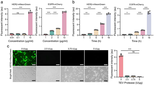
Optimization of immunostaining and fluorescence removal. (a) Fluorescent intensity as a function of NFPF concentration used for immunostaining at 4 °C (n = 5). (b) Fluorescent intensity as a function of labeling time at 4 °C (n = 5). (c) Representative fluorescence imaging and corresponding quantitative analysis of Capan-1 cells subjected to TEV protease cleavage at 30 °C for 2 h (n = 5). ns: no significant, *p < 0.05, **p < 0.01, and ***p < 0.001.
Fig. 3
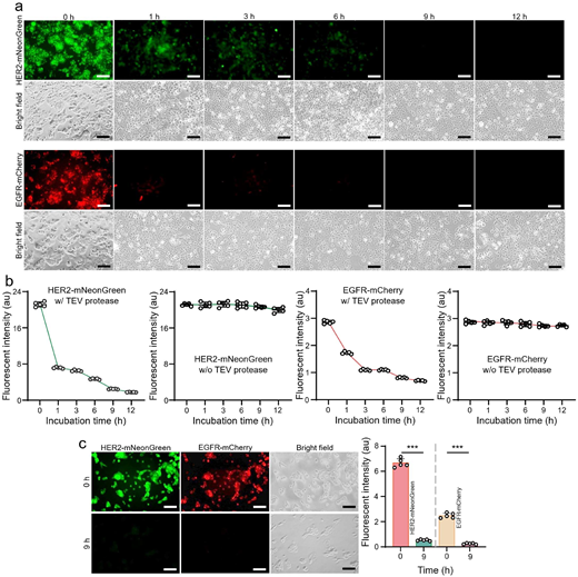
Optimization and validation of TEV-mediated fluorescence removal. (a and b) Representative fluorescence and bright-field images, together with quantitative time-course analysis, of Capan-1 cells stained with HER2-mNeonGreen or EGFR-mCherry and subjected to TEV protease cleavage at 4 °C for the indicated times. In control group, fluorescence intensity measured at six time points under conditions identical to those used in the experimental groups, except in the absence of TEV protease. Each point represents one field of view from one independently processed well (n = 5). (c) Representative images and quantitative analysis of Capan-1 cells co-labeled with HER2-mNeonGreen and EGFR-mCherry before and after TEV protease treatment at 4 °C for 9 h (n = 5). Scale bars: 500 μm. ns: no significant, *P < 0.05, and ***P < 0.001.
3.3. Iterative immunostaining
We designed an iterative labeling workflow in which distinct fluorescence signals were sequentially generated and erased through alternating cycles of NFPF staining and TEV protease treatment (Fig. 4a). Capan-1 cells were first labeled with HER2-mNeonGreen, followed by TEV protease treatment at 2.5 U μg−1 and 4 °C for 9 h to erase the green fluorescence signal. The same cells were then relabeled with EGFR-mCherry, producing red fluorescence that was subsequently removed by a second TEV protease treatment. The workflow was also performed in the reverse order, beginning with EGFR-mCherry and followed by HER2-mNeonGreen (Fig. 4b). In both sequences, fluorescence signals were sequentially generated and erased, demonstrating that iterative receptor interrogation is independent of labeling order. Importantly, the ability to relabel cells after protease treatment indicates that TEV-mediated cleavage selectively removes the fluorescent reporter without compromising receptor accessibility for subsequent immunostaining. Collectively, these results demonstrate that protease-cleavable NFPFs enable reversible fluorescence labeling and sequential imaging within the same sample.
Fig. 4
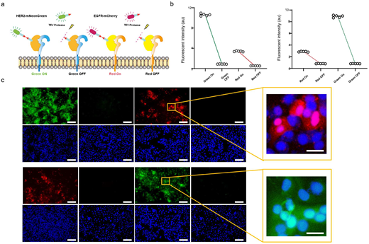
Validation of iterative immunostaining and TEV-mediated fluorescence removal. (a) Schematic of sequential HER2-mNeonGreen and EGFR-mCherry staining with TEV-mediated reporter removal. (b) Fluorescence intensity before and after each cleavage step for both staining orders. Each point represents one field from one independently processed well (n = 5). (c) Representative fluorescence and Hoechst nuclear-reference images. Images within the same fluorescence channel were displayed using identical intensity ranges. Scale bars: 500 μm. Dashed boxes indicate enlarged regions shown on the right. Scale bars: 10 μm. Representative images were obtained from corresponding experimental wells and do not necessarily depict the identical microscopic field across successive stages.
3.4. Discussion
In this study, we developed a protease-cleavable NFPF platform that enables iterative immunostaining and imaging. Our NFPF design integrates the target-recognition nanobody, TEV-cleavable peptide sequence, and fluorescent protein into a single genetically encoded fusion reagent. These NFPFs can be readily produced in cell factories, rendering our chemical-reaction-free preparation simpler, with pure NFPFs obtained via a single SEC step.28 Instead of relying on harsh chemical removal of antibodies or the destruction of fluorescent dyes, our method selectively removes the fluorescent protein domain through site-specific enzymatic cleavage, thereby erasing the fluorescence signal and facilitating subsequent rounds of immunostaining. In contrast, strategies relying on the chemical conjugation of fluorophores or DNA oligonucleotides to antibodies inevitably require multi-step processing to yield the final product, making the production workflow laborious and cost-intensive.29,30 Crucially, our NFPF platform features a highly modular, plug-and-play architecture, allowing both the NB moiety and the fluorescent protein domain to be independently and seamlessly exchanged. By pairing diverse target-specific NBs with an array of fluorescent proteins, such as mNeonGreen, mCherry, mClover3, mScarlet-I, or mCardinal, users can customize orthogonal NFPF panels tailored to specific iterative imaging requirements within a single specimen. The current results demonstrate the feasibility of this format for HER2- and EGFR-directed sequential immunostaining. Further extension to additional targets, repeated staining cycles, and improved control of fusion-protein homogeneity will be important for evaluating its broader applicability in multiplex fluorescence imaging. Notably, towards future scale-up and translational development, we plan to establish a stable HEK293 cell strain expressing the NFPFs. This stable expression system will support continuous and more standardized NFPF production while reducing variability associated with repeated transient transfections.
Considering the challenges related to yield and protein design, we selected NB as the engineered object. NB is the smallest natural antigen-binding fragment with only one immunoglobulin domain.31,32 NB exhibits remarkable stability in comparison to single-chain variable fragments (scFv) and fragment antigen-binding agents. NB can repeatedly unfold and refold without significant aggregation.33 Meanwhile, NB can retain high specificity and affinity in the nanomolar or even picomolar range under thermal and pH stress. Moreover, NB has low toxicity and low immunogenicity due to the high sequence similarity and structural homology with the human VH gene family.34 The high yield, low cost, and excellent stability of NFPFs render their preparation efficient and scalable. Notably, the anti-HER2 (2Rs15d) and anti-EGFR (7D12) NB sequences used in this study have been previously reported and are commercially available.35,36 However, because their dissociation half-lives are relatively short, it is critical to perform rinsing rapidly post-staining and proceed immediately to imaging. To afford greater operational flexibility, utilizing NBs with dissociation half-lives spanning several hours would be highly advantageous. On the other hand, given their relatively rapid dissociation kinetics, a high concentration of NFPFs was used for immunostaining. Maximizing the concentration of the NFPFs drives the binding equilibrium toward complete receptor saturation, thereby compensating for the rapid off-rate and securing sufficient initial fluorescence intensity.
We incorporated a flexible G4S linker rather than a rigid EAAAK linker.37 The G4S linker grants spatial autonomy to both the NB and the fluorescent protein, preserving high-affinity target recognition by minimizing steric hindrance. Moreover, conformational flexibility enhances the steric accessibility of the embedded cleavage motif, allowing the TEV protease to efficiently capture and process the substrate. Note that this flexible design has potential trade-offs. The linker may allow the fluorescent protein to wobble within a localized radius, potentially causing motion-induced blur in super-resolution imaging.38 Since our NFPF platform was developed for common fluorescence microscopy, this concern does not impact the current study. Additionally, flexible linkers may increase sensitivity to endogenous proteases, rendering the peptide backbone bridging the NB and fluorescent protein domains prone to spontaneous degradation.39 Our SDS-PAGE results indeed indicated partial degradation in specific NFPF samples.
TEV protease was used to facilitate cleavage of the construct. The enzyme recognizes ENLYFQG and cleaves between the Q and G residues.40 TEV protease utilizes a conserved His–Asp–Cys catalytic triad.41 The catalytic cysteine initiates nucleophilic attack on the substrate carbonyl carbon, generating a transient acyl-enzyme intermediate that is subsequently hydrolyzed to release the cleaved products. Owing to its exceptional sequence specificity, TEV protease minimizes off-target proteolysis and is widely used for recombinant protein processing. Moreover, TEV protease remains active at 4 °C, making it suitable for the processing of temperature-sensitive targets.42 Our results demonstrate that fluorescence signals can be efficiently erased without compromising cellular morphology or receptor accessibility for subsequent immunostaining. Unlike conventional methods, which rely on the removal or bleaching of fluorescent antibodies, NFPFs decouple signal elimination from target perturbation through selective proteolytic removal of the fluorescent reporter. As a result, the underlying antigenic landscape remains intact, as evidenced by the successful iterative labeling of HER2 and EGFR following TEV protease treatment. Admittedly, the catalytic kinetics of TEV protease are attenuated at 4 °C due to fundamental thermodynamic constraints.43,44 To further accelerate imaging workflows that strictly demand low-temperature handling, future iterations of this platform could substitute the TEV recognition motif with that of Human Rhinovirus (HRV) 3C protease. Known for its exceptional catalytic robustness at 4 °C, HRV 3C would provide effective cleavage without compromising the supreme sequence specificity required to prevent off-target tissue degradation.45
4. Conclusions
In this work, we developed protease-cleavable NFPFs as a reversible platform for iterative immunostaining. Incorporation of a TEV protease cleavage site enabled efficient fluorescence removal under mild conditions while preserving target accessibility and sample integrity. This genetically encoded, modular strategy avoids post-synthetic chemical conjugation and can be readily adapted to diverse nanobodies and fluorescent proteins, providing a versatile foundation for next-generation reversible imaging probes. Future efforts will focus on expanding the NFPF platform to additional molecular targets, identifying proteases with improved cleavage efficiency, and developing NFPFs optimized for in vivo molecular imaging.
Author contributions
The project was designed by YW. XX, ZK, ZS, and CC performed the experiments, collected, and analyzed the data. All authors contributed to the writing of the manuscript, discussed the results and implications, and edited the paper at all stages.
Conflicts of interest
The authors declare no competing interest.
Data availability
The authors declare that all data supporting the findings of this study are available within the paper. Source data is provided in this paper. Additional information and unique biological materials can be requested from the corresponding author upon reasonable request. Custom code was not involved in this study.
Table S1 molecular architecture of NFPFs. Fig. S1 Cell staining with free fluorescent proteins. Fig. S2 Photobleaching assessment of fluorescent proteins. Supplementary information (SI) is available. See DOI: https://doi.org/10.1039/d6an01102k.
Acknowledgements
This work was partially supported by the National Cancer Institute (R37CA255948).
References
- E.von Witting,S.Hober and S.Kanje,Affinity-Based Methods for Site-Specific Conjugation of Antibodies,Bioconjugate Chem.,2021,32,1515–1524.
- S.Vira,E.Mekhedov,G.Humphrey and P. S.Blank,Fluorescent-labeled antibodies: Balancing functionality and degree of labeling,Anal. Biochem.,2010,402,146–150.
- Á.Szabó, et al.,The Effect of Fluorophore Conjugation on Antibody Affinity and the Photophysical Properties of Dyes,Biophys. J.,2018,114,688–700.
- A.Beck,L.Goetsch,C.Dumontet and N.Corvaïa,Strategies and challenges for the next generation of antibody–drug conjugates,Nat. Rev. Drug Discovery,2017,16,315–337.
- M. J.Gerdes, et al.,Highly multiplexed single-cell analysis of formalin-fixed, paraffin-embedded cancer tissue,Proc. Natl. Acad. Sci. U. S. A.,2013,110,11982–11987.
- J.Kennedy-Darling, et al.,Highly multiplexed tissue imaging using repeated oligonucleotide exchange reaction,Eur. J. Immunol.,2021,51,1262–1277.
- Y.Goltsev, et al.,Deep Profiling of Mouse Splenic Architecture with CODEX Multiplexed Imaging,Cell,2018,174,968–981.
- R.Jungmann, et al.Multiplexed 3D cellular super-resolution imaging with DNA-PAINT and Exchange-PAINT,Nat. Methods,2014,11,313–318.
- S.Black, et al.,CODEX multiplexed tissue imaging with DNA-conjugated antibodies,Nat. Protoc.,2021,16,3802–3835.
- R.Jungmann, et al.,Multiplexed 3D Cellular Super-Resolution Imaging with DNA-PAINT and Exchange-PAINT,Nat. Methods,2014,11,313–318.
- F.Ji, et al.,Multiplex Protein Imaging through PACIFIC: Photoactive Immunofluorescence with Iterative Cleavage,ACS Bio Med Chem Au,2023,3,283–294.
- N.Mougios, et al.,NanoPlex: a universal strategy for fluorescence microscopy multiplexing using nanobodies with erasable signals,Nat. Commun.,2024,15,8771.
-
F.Ji, et al.,Multiplex Protein Imaging through PACIFIC: Photoactive Immunofluorescence with Iterative Cleavage,ACS Bio Med Chem Au,2023,3,283–294.
-
M.Mondal,R.Liao,L.Xiao,T.Eno and J.Guo,Highly Multiplexed Single-Cell In Situ Protein Analysis with Cleavable Fluorescent Antibodies,Angew. Chem., Int. Ed.,2017,56,2636–2639.
-
I.Vaneycken, et al.,Preclinical screening of anti-HER2 nanobodies for molecular imaging of breast cancer,FASEB J.,2011,25,2433–2446.
-
A.Noor, et al.,Production of a mono-biotinylated EGFR nanobody in the E. coli periplasm using the pET22b vector,BMC Res. Notes,2018,11,751.
-
M.Arslan, et al.,Effect of non-repetitive linker on in vitro and in vivo properties of an anti-VEGF scFv,Sci. Rep.,2022,12,5449.
-
P.Kunz, et al.,The structural basis of nanobody unfolding reversibility and thermoresistance,Sci. Rep.,2018,8,7934.
-
X.Wang, et al.,A designed fusion tag improves soluble expression of nanobody in E. coli,Magn. Reson. Lett.,2026,200287https://doi.org/10.1016/j.mrl.2026.200287.
-
Frontiers|Recombinant protein expression in Escherichia coli: advances and challenges. https://www.frontiersin.org/journals/microbiology/articles/10.3389/fmicb.2014.00172/full.
-
X.Xi, et al.,A comparative study and evaluation of anti-EGFR nanobodies expressed in Pichia pastoris and Escherichia coli as antitumor moieties,Protein Expression Purif.,2021,184,105888.
-
I.Vaneycken, et al.,Preclinical screening of anti-HER2 nanobodies for molecular imaging of breast cancer,FASEB J.,2011,25,2433–2446.
-
D.Lee and G.Lee,Single-molecule studies of repair proteins in base excision repair,BMB Rep.,2025,58,17–23.
-
M.Shahriari,F.Shafiee,F.Moazen and H. M.Mohammad Sadeghi,Optimization of the Production of Soluble Recombinant TEV Protease in Two E. coli Strains,Avicenna J. Med. Biotechnol.,2024,16,279–283.
-
A. S.Chaudhry,Comparing two commercial enzymes to estimate in vitro proteolysis of purified or semi-purified proteins,J. Anim. Physiol. Anim. Nutr.,2005,89,403–412.
-
E.Buck, et al.,Inactivation of Akt by the epidermal growth factor receptor inhibitor erlotinib is mediated by HER-3 in pancreatic and colorectal tumor cell lines and contributes to erlotinib sensitivity,Mol. Cancer Ther.,2006,5,2051–2059.
-
C. G.England,R.Hernandez,S. B. Z.Eddine and W.Cai,Molecular Imaging of Pancreatic Cancer with Antibodies,Mol. Pharm.,2016,13,8–24.
-
N.Mougios, et al.,NanoPlex: a universal strategy for fluorescence microscopy multiplexing using nanobodies with erasable signals,Nat. Commun.,2024,15,8771.
-
J.Wiener,D.Kokotek,S.Rosowski,H.Lickert and M.Meier,Preparation of single- and double-oligonucleotide antibody conjugates and their application for protein analytics,Sci. Rep.,2020,10,1457.
-
T.Wang, et al.,Constructions, Purifications and Applications of DNA-Antibody Conjugates: A Review,ACS Omega,2024,9,47951–47963.
-
J.Liu, et al.,Unveiling the new chapter in nanobody engineering: advances in traditional construction and AI-driven optimization,J. Nanobiotechnol.,2025,23,87.
-
T. D.Meyer,S.Muyldermans and A.Depicker,Nanobody-based products as research and diagnostic tools,Trends Biotechnol.,2014,32,263–270.
-
A.de Marco,Recombinant expression of nanobodies and nanobody-derived immunoreagents,Protein Expression Purif.,2020,172,105645.
-
N. E.Eskier,D.Eskier,E.Firuzan and S. K.Uzunlar,Physicochemical differences between camelid single-domain antibodies and mammalian antibodies,Turk. J. Biol.,2023,47,423–436.
-
M.Ducharme, et al.,Evaluation of [89Zr]Zr-DFO-2Rs15d Nanobody for Imaging of HER2-Positive Breast Cancer,Mol. Pharm.,2023,20,4629–4639.
-
K. R.Schmitz,A.Bagchi,R. C.Roovers,P. M. P.van Bergen en Henegouwen and K. M.Ferguson,Structural Evaluation of EGFR Inhibition Mechanisms for Nanobodies/VHH Domains,Structure,2013,21,1214–1224.
-
Z.Huang,G.Li,C.Zhang and X.-H.Xing,A study on the effects of linker flexibility on acid phosphatase PhoC-GFP fusion protein using a novel linker library,Enzyme Microb. Technol.,2016,83,1–6.
-
J.Wang, et al.,MINFLUX reveals nonnormal distribution of linkage errors in immunofluorescence labeling,Commun. Phys.,2025,8,253.
-
Z.Liu, et al.,Peptidoglycan-Targeting Staphylolytic Enzyme Lysostaphin as a Novel and Efficient Protease toward Glycine-Rich Flexible Peptide Linkers,J. Agric. Food Chem.,2023,71,5293–5301.
-
R. B.Kapust, et al.,Tobacco etch virus protease: mechanism of autolysis and rational design of stable mutants with wild-type catalytic proficiency,Protein Eng.,2001,14,993–1000.
-
J.Phan, et al.,Structural Basis for the Substrate Specificity of Tobacco Etch Virus Protease,J. Biol. Chem.,2002,277,50564–50572.
-
S.Raran-Kurussi,J.Tözsér,S.Cherry,J. E.Tropea and D. S.Waugh,Differential temperature dependence of tobacco etch virus and rhinovirus 3C proteases,Anal. Biochem.,2013,436,142–144.
-
V. L.Arcus, et al.,On the Temperature Dependence of Enzyme-Catalyzed Rates,Biochemistry,2016,55,1681–1688.
-
N. E.Ketaren, et al.,Unique mechanisms to increase structural stability and enhance antigen binding in nanobodies,Structure,2025,33,677–690.
-
D. S.Waugh,An overview of enzymatic reagents for the removal of affinity tags,Protein Expression Purif.,2011,80,283–293.

